Archive

  • Select all
    |
    Reviews
  • YANG Jian, LIAO Li, TIAN Wei-Dong
    Chinese Journal of Biochemistry and Molecular Biol. 2021, 37(8): 989-997. https://doi.org/10.13865/j.cnki.cjbmb.2021.03.1508
    Abstract ( ) Download PDF ( )   Knowledge map   Save
    Exosomes are phospholipid bilayer extracellular vesicles secreted by cells, and they act as carriers for material transfer and information exchange between cells. Exosomes exist in many different biofluids and have good application prospects in disease diagnosis and drug delivery. Due to the nano-scale size and heterogeneity of exosomes, as well as the complex composition of biofluids, the separation of exosomes from biofluids is particularly difficult. At present, there are six commonly used methods forisolating exosomes from biofluids: ultracentrifugation, precipitation, size exclusion chromatography, density gradient centrifugation, ultrafiltration and immuno-affinity membrane. Each method has its own advantages and disadvantages. It is difficult to separate and obtain high-quality exosomes when used alone. There are also differences in the selection of methods for separating exosomes from different biofluids. With the continuous emergence of some new technologies such as microfluidic technology and the continuous improvement of classical methods, the exosome separation technology is improved, which is beneficial to the application of exosomes in clinical and scientific research. This review compares and evaluates the isolation and identification methods of exosomes from different biofluid sources, summarizes the selection and differences of exosome isolation methods from different biofluid sources, and provides a reference for subsequent research on exosomes from biofluids.
  • NIU Xiao-Yun, XIE Xi-Xiu
    Chinese Journal of Biochemistry and Molecular Biol. 2021, 37(8): 998-1004. https://doi.org/10.13865/j.cnki.cjbmb.2021.01.1376
    Abstract ( ) Download PDF ( )   Knowledge map   Save
    Thrombomodulin (TM) is a single-chain transmembrane glycoprotein that mainly exists in vascular endothelial cells, hematopoietic progenitor cells, monocytes and macrophages. TM is mainly composed of five structural regions: the N-terminal lectin-like domain which plays a role in anti-inflammatory, and the six epidermal growth factor-like repeats which function as coagulation and fibrinolysis as well as serine-rich threonine regions and transmembrane domains and cytoplasmic domains. TM exhibits anti-inflammatory and anticoagulant effects by binding to thrombin to activate protein C, and TM-thrombin complex can also activate fibrinolytic inhibitors to suppress fibrinolysis. Previous reports showed that inhibiting epithelial mesenchymal transformation, mitogen-activated protein kinase or activating protein C and fibrinolytic inhibitor are the major mechanisms by which TM exerts anti-tumor properties. In atherosclerosis, TM can prevent atherosclerosis by blocking the activation of thrombin-mediated PAR-1 and inhibiting autophagy and apoptosis of endothelial cells. TM lectin-like domains can also bind to thrombin to inhibit its activity and further inhibit pulmonary thrombosis, fibrosis and inflammation. Moreover, TM protein is also involved in the pathogenesis of diabetic nephropathy, preeclampsia and ischemia-reperfusion injury. At present, TM is only clinically used for the treatment of sepsis and disseminated intravascular coagulation. Its role and therapeutic potential in cardiovascular and cerebrovascular diseases, cancers and other diseases deserve further exploration.
  • ZHAO Qi, MO Ri-Gen, FAN Li-Fei
    Chinese Journal of Biochemistry and Molecular Biol. 2021, 37(8): 1005-1009. https://doi.org/10.13865/j.cnki.cjbmb.2020.12.1501
    Abstract ( ) Download PDF ( )   Knowledge map   Save
    Cancer-associated fibroblasts(CAFs) are important cellular components of the tumor microenvironment. They have a variety of cellular sources, including resident fibroblasts, bone marrow mesenchymal stem cells and epithelial cells, which contribute to the development of tumors. CAFs play important roles in cancer initiation, progression, and metastasis,which can promote tumor proliferation and migration, promote tumor angiogenesis, regulate tumor immunity, and improve tumor drug resistance. Therefore, it is a promising development direction in tumor-targeted therapy. CAFs regulate the biological characteristics of tumor cells and other stromal cells through cell-to-cell contact, releases many regulatory factors and synthesizes and reshapes the extracellular matrix, and influences the occurrence and development of cancer in these ways. However, there are still many unresolved issues on the way in targeting CAFs for tumor therapy. For example, the origin and functional heterogeneity of CAFs still need to be further explored. This review mainly focuses on the summary of origin of CAFs, their roles in tumor development and the potential application in cancer targeted therapy, which will help to deepen the understanding of the roles of CAFs in cancer development and future cancer treatment.
  • CHENG Xi-Yao, LI Gang, SU Zheng-Ding
    Chinese Journal of Biochemistry and Molecular Biol. 2021, 37(8): 1010-1016. https://doi.org/10.13865/j.cnki.cjbmb.2021.04.1511
    Abstract ( ) Download PDF ( )   Knowledge map   Save
    Conformation dynamics attributes to the biological functions of active proteins and conformation ensembles. The conformation ensembles include protein stable states (PSS) that can be measured by conventional structural biology approaches and the invisible protein states (IPS) that cannot be measured by conventional structural biology ones. The conformational exchange between IPS and PSS plays an important role in the biological functions of proteins. In this review, we briefly describe the basic properties of IPS and discuss its contribution to the development of the classical molecular recognition mechanism of “key-lock hypothesis” and "induced fit hypothesis" into "conformational selection hypothesis". Furthermore, this review compares the advantages and disadvantages of the current structural biology approaches for investigating the conformational properties of IPS. Because of advanced NMR technology, the exploration of the conformational properties of IPS in experimental manner has become feasible. It is expected that the study of IPS and its function will not only help clarify the molecule recognition mechanism of proteins, but also provide a basis for guiding the design of targeted drugs.
  • WEI Qiu-Yu, LI Te-Te, CHEN Jing-Tao
    Chinese Journal of Biochemistry and Molecular Biol. 2021, 37(8): 1017-1023. https://doi.org/10.13865/j.cnki.cjbmb.2021.01.1513
    Abstract ( ) Download PDF ( )   Knowledge map   Save
    Dendritic cells (DCs) are specialized antigen-presenting cells. Immature dendritic cells can be activated into mature dendritic cells by recognizing antigens, then the antigens are processed and presented to T lymphocytes. DCs play a vital role in initiating immune response, regulating immune response and maintaining immune tolerance. Therefore, regulating the immune function of DCs can be used to treat diseases such as autoimmune diseases and tumors. With the deepening of research on the regulation of DCs, people have gradually realized that the existence of reactive oxygen species (ROS) in DCs is of great significance. ROS is a term of strong oxidizing reactive species, and the dynamic balance of its production and removal is the key to maintaining cell redox homeostasis. ROS of physiological level is an important molecule involved in a variety of signal pathways, which can regulate cell growth, differentiation and different physiological and biochemical reactions. The changes in the level of ROS affect the state and function of cells. In addition, because there are many ways to produce ROS in the cell, the effects of different sources of ROS on DCs are not usually the same; and even the same source of ROS may have different effects when cells are in different states. This article summarized the influence of intracellularROS changes and different sources of ROS on the differentiation, maturation and function of DCs, aiming to reveal how ROS plays an important role in regulating the immune function of DCs. At the same time, this article also showed the urgent need for the in-depth study of ROS regulating the function of DCs, which may help the application of DCs immune regulation to a wider range of disease treatments.
  • TAO Su-Wan, ZHU Lian, XU Na
    Chinese Journal of Biochemistry and Molecular Biol. 2021, 37(8): 1024-1031. https://doi.org/10.13865/j.cnki.cjbmb.2021.02.1492
    Abstract ( ) Download PDF ( )   Knowledge map   Save
    Skin wound healing is one of the difficult problems in clinical medicine. The long-term wound healing has a great impact on patients and causes huge social and economic burden. Hydrogels have good biocompatibility, biodegradability and plasticity, and have excellent properties of hemostasis, antibacterial and water retention. They are widely used as skin wound dressing. The exosomes derived from stem cells contain the unique contents and membrane components of stem cells. These exosomes have the similar repair function as stem cells, and avoid the safety problems of teratoma and graft immune rejection caused by stem cell therapy, and therefore has become the research hotspot of cell-free repair. However, exosomes in the treatment process will face high clearance rate, short half-life and difficulty in large-scale preparation, which limits the therapeutic effect of exosomes. Hydrogel loaded stem cell exosomes can not only prevent exosome from being rapidly removed, but also play the role of exosome and hydrogel in promoting wound healing, skin repair synergy, and thereafter has potential application value in wound repair research. In this paper, production and characteristics of exosomes, commonly used hydrogel materials, stem cell exosomes from different sources, exosomes extraction and identification methods have been summarized. Furthermore, stem cell exosomes related mechanisms of wound repair, protective and sustained release effects of hydrogel on exosomes, and synergistic effects of hydrogel and exosome are also reviewed. This paper provides a reference for the future research in the field of skin repair.
  • Research Papers
  • LIU Si, WANG Fang, PEI Sheng-Qiang, PEI Jian-Qiu, CONG Xiang-Feng, CHEN Xi
    Chinese Journal of Biochemistry and Molecular Biol. 2021, 37(8): 1032-1039. https://doi.org/10.13865/j.cnki.cjbmb.2021.06.1270
    Abstract ( ) Download PDF ( )   Knowledge map   Save
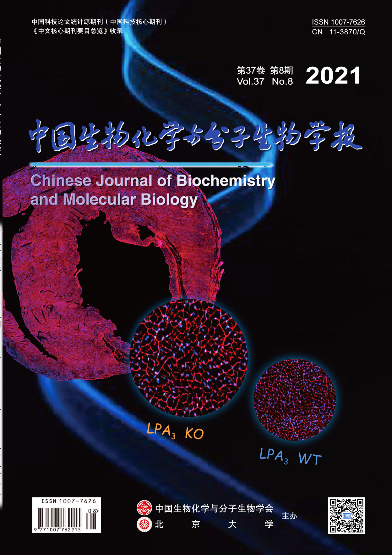
    Lysophosphatidic acid(LPA)is a small bioactive phospholipid that mediates various cellular processes such as proliferation, survival, and migration. In particular, LPA signaling has been shown to affect the development of diverse tissues. Our previous work demonstrated that LPA could promote primary neonatal rat cardiomyocytes proliferation. However, the role of LPA and its receptor in postnatal heart development is unknown. By using databases for biological information and RT-qPCR, we analyzed the expression of six LPA receptors (LPA1-LPA6) and found that LPA3 was the most highly expressed LPA receptor in human and mouse hearts. In addition, the expression of LPA3 mRNA at P7 peaked at 26-fold higher than adults’ expression. Using LPA3-knockout mice, we found that the number of proliferating cardiomyocytes, detected by co-immunostaining pH3 with cardiac troponin T, was significantly decreased in the LPA3-knockout mice during the first week of postnatal life (P<0.05). Furthermore, by using cardiac echocardiography and pathological staining, we found that LPA3-knockout adult mice displayed normal cardiac morphology and function. However, the cardiomyocytes volume increased compensatorily (P<0.05). In addition, using a myocardial infarction (MI) model, we found that the cardiac function decreased and the infracted area increased after myocardial infarction at P7 (P<0.05). In summary, our study reported that LPA3-mediated LPA signaling was a crucial factor for heart development in the postnatal heart, which suggested that LPA3 might be a potential drug target for the treatment of the myocardial injury.
  • YANG Bin, SONG Ge, SHI Li-Ying, TANG Hua
    Chinese Journal of Biochemistry and Molecular Biol. 2021, 37(8): 1040-1049. https://doi.org/10.13865/j.cnki.cjbmb.2021.05.1066
    Abstract ( ) Download PDF ( )   Knowledge map   Save
    Circular RNAs play an important role in tumorigenesis. In this study, we explored the role of aberrant expression of circHMGCS1 in cervical cancer cells with TNF-α treatment. RT-qPCR assays showed that circHMGCS1 was highly expressed after TNF-α treatment. And then we investigated the cyclization and stabilization of circHMGCS1 by RNase R and half-life analysis. We further revealed that overexpression of circHMGCS1 promoted the malignancy of cervical cancer cells and the expression of the epithelial-mesenchymal transition (EMT) markers by Transwell analysis, MTT, clone formation and Western blotting. Furthermore, we found that circHMGCS1 activated the NF-κB signaling pathway. Altogether, this study provides an experimental basis for elucidating the role of circHMGCS1 in cervical cancer cells, and provides a potential biomarker for the diagnosis and treatment of cervical cancer.
  • DONG Chun-Yang, WANG Bo, MING Jin, LI Chen, FANG Shi-Cai, HUANG Yi, LIU Jing
    Chinese Journal of Biochemistry and Molecular Biol. 2021, 37(8): 1050-1061. https://doi.org/10.13865/j.cnki.cjbmb.2021.06.1084
    Abstract ( ) Download PDF ( )   Knowledge map   Save
    Embryonic stem cells (ESCs) have the ability to differentiate into various adult cells, and their fate in the process of development and differentiation is determined by the comprehensive regulation of multiple factors such as gene expression, epigenetics, and extracellular signals. Epigenetic regulation, such as DNA methylation, histone acetylation, and methylation, plays an important role in the maintenance of pluripotency and differentiation of ESCs. Suds3 (Sin3 histone deacetylase corepressor complex component SDS3) is one of the important components of Sin3 histone deacetylase complex. It played an important role in embryonic development, cell proliferation, chromosome separation and other biological processes. However, the functions of Suds3 in ESCs, such as its influence on the proliferation, maintenance of pluripotency and differentiation of ESCs, were rarely reported. In this study, we used CRISPR/Cas9 gene editing technology to construct a Suds3 knockout mouse embryonic stem cell line, and combined cell culture, in vitro embryoid body (EB) formation and in vivo teratoma formation, CCK-8 and cell counting experiments to study the function of Suds3 in ESCs. Western blotting results showed that the SUDS3 protein was not expressed, and the Suds3 gene was successfully knocked out. Through the observation of cell morphology and fluorescence quantitative PCR (QRT-PCR) to detect the expression of pluripotency genes, we found that the knockout of Suds3 had no significant effect on the maintenance of pluripotency of ESCs. Embryoid body (EB) formation experiments revealed that on the fourth and sixth days of EB formation, the pluripotency gene expression was not down-regulated as quickly as WT cells but increased in Suds3-/- cells (***P<0.001), the gene expression of mesoderm and endoderm was significantly lower than that of wild type (***P<0.001), but the expression level of ectoderm genes is higher than that of wild type (***P<0.001). The results of CCK8 proliferation experiment and cell counting experiment found that the knockout of Suds3 inhibited the proliferation ability of ESCs (*P<0.05,***P<0.001). In vivo teratoma formation experiments and HE staining showed that Suds3 knockout inhibited proliferation, at the same time promoted ectoderm differentiation, and reduced mesoderm and endoderm differentiation. And overexpression of Suds3 in Suds3-/- cells can rescue the phenotypic changes of ESCs. In sum, our experiment successfully constructed a Suds3 knockout embryonic stem cell line, and showed that the knockout of Suds3 had no significant effect on the maintenance pluripotency, but inhibited the proliferation of ESCs and promoted the differentiation of ectoderm, and limited the differentiation of mesoderm and endoderm. The results above provide new insights and research models for studying the effects of histone acetylation on the differentiation and early embryonic development of ESCs, and provide a technical reference for in vitro targeted differentiation to obtain functional cells and future cell therapy strategies of embryonic stem cells.
  • WANG Ling-Yan, WANG Yu-Han, ZHANG Cheng, CHEN Xi-Xi, HUANG Huang, LI Wen-Li, ZHANG Jia-Ning, LIU Yu-Bo
    Chinese Journal of Biochemistry and Molecular Biol. 2021, 37(8): 1062-1068. https://doi.org/10.13865/j.cnki.cjbmb.2021.05.1024
    Abstract ( ) Download PDF ( )   Knowledge map   Save
    Caveolin-1 (Cav-1), a major structural protein of caveolae, is implicated in the vesicular uptake processes of transcytosis and cell signaling. However, its role in modulating protein glycosylation and tumor metastasis remains to be further elucidated. In the present study, it was shown that Cav-1 promotes the expression of O-GlcNAcylation and O-GlcNAc transferase (OGT), and triggers the invasion and metastasis of hepatocellular carcinoma (HCC) cells. The results of RT-qPCR, Western blot and dual luciferase reporter assay showed that Cav-1 negatively regulated the expression of transcription factor RUNX2 in HCC. Subsequently, this results in attenuate RUNX2-induced transcription of miR24. miR24 suppresses mouse HCC cells invasion and metastasis via directly targeting Ogt mRNA 3′UTR. This research provides evidence of Cav-1-mediated OGT expression and O-GlcNAc (O-linked N-acetylglucosamine) elevation. These data give insight into a novel mechanism of HCC occurrence and development.
  • YAN Shi-Ke, FENG Jing-Wen, CHANG Jiao, ZHANG Bing, DUAN Na-Min, WU Wen-Hui, LIU Ning
    Chinese Journal of Biochemistry and Molecular Biol. 2021, 37(8): 1069-1077. https://doi.org/10.13865/j.cnki.cjbmb.2021.05.1049
    Abstract ( ) Download PDF ( )   Knowledge map   Save
    FGFC1 (Fungi fibrinolytic compound1) is a bisindole compound with good biological activity, which was first derived from the Stachybotrys longispora FG216. However, the anti-tumor effects of FGFC1 have not been reported. This study investigated the effect and mechanism of FGFC1 on the proliferation, apoptosis, migration and invasion of non-small cell lung cancer (NSCLC) cells.Firstly, PC9, H1975, HCT116, HeLa and 293T cells were treated with different concentrations of FGFC1, and the cell counting kit-8 assay was used to determine relative cell viability; flow cytometry was used to evaluate apoptosis; real-time PCR and Western blotting analysis were performed to measure the expression of apoptosis-related genes in PC9 cells; wound healing and Transwell invasion assays were used to measure the ability of migration and invasion; Western blotting was performed to measure the expression of kinase proteins involved in the PI3K/Akt/mTOR signaling pathway, exploring the influence of FGFC1 on this signaling pathway. We found that FGFC1 selectively inhibited the proliferation of PC9 cells. It also up-regulated the expression of apoptosis-promoting protein cleaved-caspase-3 and cleaved-PARP, and induced apoptosis in a dose-dependent manner (P < 0.05). FGFC1 also significantly inhibited the migratory and invasive capacity of PC9 cells in a dose-dependent manner (P < 0.05). Further studies confirmed that FGFC1 could inhibit the activation of the PI3K/Akt/mTOR signaling pathway with the down-regulation of the protein expression levels ofp-PI3K, p-Akt and p-mTOR. Thus, we conclude that FGFC1 inhibited the proliferation of PC9 and H1975 cells, induced the apoptosis and inhibited the migration and invasion of PC9 cells, which may take place through down-regulating the PI3K/Akt/mTOR signaling pathway. These findings suggest that FGFC1 might be a new therapeutic target in NSCLC treatment in the future.
  • HUANG Yuan-Feng, LI Pan, YANG Ming-Qiang, LIU Hao, CHEN Dan-Yang
    Chinese Journal of Biochemistry and Molecular Biol. 2021, 37(8): 1078-1084. https://doi.org/10.13865/j.cnki.cjbmb.2021.06.1215
    Abstract ( ) Download PDF ( )   Knowledge map   Save
    EGFR tyrosine kinase inhibitor (EGFR-TKI)-targeted therapy has been playing an important role in the treatment of non-small cell lung cancer (NSCLC). However, unavoidable therapeutic resistance significantly limits the clinical efficacy of TKI. As an important member of the PAK family of serine/threonine kinases, p21-activated protein kinase 2 (PAK2) plays a critical role in tumorigenesis and tumor development. The aim of this work is to investigate the effect and molecular mechanism of inhibition of PAK2 on the reversal of gefitinib resistance in NSCLC. Firstly, Western blotting was used to detect the expression and phosphorylation level of PAK2 in gefitinib-resistant HCC827/GR cells. The results showed that the phosphorylation level of PAK2 was significantly increased in HCC827/GR cells compared with HCC827 cells (P < 0.01), while the total protein level of PAK2 did not change. Then, HCC827/GR cells were treated with PAK2 inhibitors FRAX597 or G5555. The MTS assay and clone formation assay results showed that FRAX597 or G5555 significantly increased the sensitivity of HCC827/GR cells to gefitinib (P < 0.01). Flow cytometry analysis showed that treatment of FRAX597 could induce cell cycle arrest in G2/M phase of HCC827/GR cells, and increased gefitinib-induced apoptosis and cleaved Caspase-3 expression (P < 0.01). Western blotting results showed that treatment of FRAX597 significantly inhibited the expression of BCL-2 and CDK4 (P < 0.05), but increased the expression of BAX, p21 and p27 (P < 0.05). Taken together, these results indicated that activation of PAK2 is closely related to NSCLC with gefitinib resistance. Inhibition of PAK2 could induce cell cycle arrest and cell apoptosis, which increased the sensitivity of NSCLC cells to gefitinib.
  • TANG Qing-Hua, SUN Xue-Hui, LI Hong-Li, HU Wen-Ting, HU Kai-Li, FAN Xin
    Chinese Journal of Biochemistry and Molecular Biol. 2021, 37(8): 1085-1092. https://doi.org/10.13865/j.cnki.cjbmb.2021.05.1018
    Abstract ( ) Download PDF ( )   Knowledge map   Save
    Tongue squamous cell carcinoma (TSCC) is the most common type of oral squamous cell carcinoma (OSCC) with high morbidity and mortality. Many studies have shown that microRNA (miRNA) are small non-coding RNA that regulate the post-transcriptional processing of target genes, resulting in the degradation and translation inhibition of target mRNA. However, how the transmembrane p24 trafficking protein 2 (TMED2) is regulated by miR-5583-5p on migration, invasion, proliferation and epithelial-mesenchymal transition (EMT) of TSCC Cal-27 cells is unclear. In this study, a database was used to analyze the expression of TMED2 in HNSCC (P <0.001) in head and neck cancer (HNC). Western blot showed that the expression of TMED2 protein was up-regulated in 6 cases of TSCC tissues and cell lines such as SCC-9, SCC-25 and CAL-27. After the Cal-27 cells transfected with TMED2 interference plasmid (SiTMED2) the expression of E-cadherin was up-regulated, and N-cadherin and Vimentin was down-regulated. Migration and invasion experiments showed that the number of cells transfused into the basement membrane of the cells was lower than that of the control group (P<0.05). The results of EdU showed that the proliferation of Cal-27 cells transfected with SiTMED2 was decreased (P<0.05). The results of dual luciferase experiment showed that TMED2 had a binding target to miR-5583-5p, and the expression of miR-5583-5p in Cal-27 cell was lower than that in Hoec cells. The expression of miR-5583-5p was increased and TMED2 protein was decreased after the Cal-27 cells were transfected with miR-5583-5p plasmid (P < 0.05). In conclusion, TMED2 is regulated by miR-5583-5p and promoted the migration, invasion, proliferation and EMT of tongue squamous cell carcinoma cell Cal-27.
  • YANG Wei-Yu, LIU Chi, XIE Hui, LIN Pin, YU Xiao-Guang
    Chinese Journal of Biochemistry and Molecular Biol. 2021, 37(8): 1093-1099. https://doi.org/10.13865/j.cnki.cjbmb.2021.06.1629
    Abstract ( ) Download PDF ( )   Knowledge map   Save
    Prostate cancer is a common malignant tumor in male patients. It is of great clinical significance to explore the pathogenesis of prostate cancer and find suitable therapeutic targets. NR4A3 is derived from the nuclear hormone receptor superfamily of steroids, and NR4A3 plays an important role in the malignant progression of a variety of tumors. However, its role in prostate cancer has not yet been elucidated. Therefore, this project intends to investigate the role of NR4A3 in prostate cancer and screen for miRNAs that target NR4A3, which may help find potential target for the diagnosis and treatment of prostate cancer. The GEPIA website predicts that NR4A3 is under-expressed in prostate cancer tissues, and qRT-PCR data confirmed downregulation of NR4A3 in prostate cancer cells (P<0.01). CCK8 and clone formation experiments show that overexpression of NR4A3 can significantly inhibit the viability, the number and size of colonies of prostate cancer cells (P<0.01). The bioinformatics website predicts that NR4A3 may be the target gene of miR-20a, and qRT-PCR showed that miR-20a expression was elevated in prostate cancer cells (P<0.01). Furthermore, dual luciferase reporter gene experiment confirmed that miR-20a can target two sites of 3′-UTR of NR4A3 (P<0.05, P<0.001). Western blot results showed that miR-20a can inhibit the expression of NR4A3. CCK8 experiments further found that miR-20a inhibitor can significantly reduce the viability of prostate cancer cells(P<0.05), while miR-20a mimic has the opposite effect (P<0.05, P<0.01). CCK8 and clone formation experiments showed that when co-transfected with miR-20a mimic and pcDNA3.1-NR4A3 recombinant plasmids, up-regulation of NR4A3 could partially offset the viability, the number and size of colonies of PC3 cells promoted by miR-20a mimic (P<0.05) . In summary, miR-20a promotes the proliferation of prostate cancer cells by targeting NR4A3.
  • SUN Zhong-Xing, GE Ke-Li, ZHANG Zhi-Xia, JIA Shao-Ping, ZHANG Jin-Yu, GE Yin-Lin
    Chinese Journal of Biochemistry and Molecular Biol. 2021, 37(8): 1100-1109. https://doi.org/10.13865/j.cnki.cjbmb.2021.05.10663
    Abstract ( ) Download PDF ( )   Knowledge map   Save
    Hyperuricemia is a chronic metabolic disease caused by the accumulation of uric acid in the body caused by purine metabolism disorder. In recent years, the incidence of hyperuricemia has increased and the age of onset is showing a younger trend. Finding effective therapeutic targets and treatment methods is a hot spot of current research. The urate transporter ATP-binding cassette subfamily G member 2 (ABCG2) is mainly expressed in the kidney and promotes uric acid excretion. In this study, ABCG2 mRNA was synthesized in vitro and transfected into hyperuricemia model mice to observe its effect on mouse uric acid levels. Firstly, the DNA template of ABCG2 mRNA was chemically synthesized, and then transcribed into mRNA in vitro, followed by modification and transfection into mouse TCMK-1 renal tubular epithelial cells. Finally, the protein expression in the cells was detected by Western blot. The results showed that the amount of protein expression in TCMK-1 cells was positively correlated with the amount of transfected mRNA (P<0.01), indicating a successful transfection. In animal experiments, twenty-four SPF mice were randomly divided into four groups (n=6): control group, hyperuricemia model group, benzbromarone group [20 mg/(kg·d)] and mRNA group [2 mg/(kg·3d)]. The mice have been modeled and treated for 28 days. During this period, the body weight and growth status of the mice were monitored daily. After the treatment, the levels of serum uric acid, urine uric acid, serum creatinine, blood urea nitrogen and liver xanthine oxidase were analyzed. The results showed that compared with the model group of mice, mRNA treatment can significantly reduce the levels of serum uric acid (100.38 ± 10.94), blood urea nitrogen (6.30 ± 1.10), and serum creatinine (30.86 ± 5.78,P<0.05 or P<0.01). It can also increase the level of urine uric acid (617.48 ± 50.34,P<0.05) in mice and promote the excretion of uric acid. But it has no significant effect on the activity of xanthine oxidase (26.19 ± 2.58) in the liver. The pathological changes of mice kidney were observed by HE staining. The results showed that compared with mice in the model group, pathological damages such as renal tubular cell edema and inflammatory cell infiltration in the mRNA treatment group were significantly improved. The relative expression of mRNA in mice kidney was detected by qRT-PCR, and the protein expression of ABCG2 in mice kidney was detected by immunohistochemistry and Western blot. The results showed that the relative expression of ABCG2 mRNA and its protein were significantly up-regulated in the kidney tissues of mice in the mRNA group (P < 0.01), indicating that the transfection was successful in vivo. In conclusion, ABCG2 mRNA synthetized and modified in vitro can be successfully expressed in hyperuricemia mice and promote excretion of uric acid and other organic ions, as well as improvement of renal injury in mice. These results provide experimental basis for the clinical application of ABCG2 as a target for the treatment of hyperuricemia related diseases.
  • FU Peng-Yu, JIA Jie, ZHU Rong-Xin, GU Bo-Ya, GONG Li-Jing
    Chinese Journal of Biochemistry and Molecular Biol. 2021, 37(8): 1110-1117. https://doi.org/10.13865/j.cnki.cjbmb.2021.05.1689
    Abstract ( ) Download PDF ( )   Knowledge map   Save
    The high altitude/hypoxic environment induced skeletal muscle atrophy is considered to be the interaction of multi-system and multi-organ, but the direct mechanism of hypoxia on muscle cells in this process is not clear. This study intended to investigate the effects of hypoxia exposure on proteins in ubiquitin and autophagy pathways, and explored the possible mechanism of hypoxia induced change of myotube diameter. The expression of myosin, hypoxia inducible factor-1 α (HIF-1α), forkhead box protein O1 (FoxO1), and ubiquitin protease pathway (MuRF1 and Atrogin1) and autophagy lysosomal pathway (p62, Beclin1, LC3) related proteins were detected by Western blot; The integrated optical density (IOD) of Myosin and LC3 was detected by IF. The results showed that the diameters of myotube at 6 h and 12 h were significantly reduced, and the expression of myosin was significantly reduced at 6 h after hypoxia exposure (P<0.05); the protein levels of HIF-1α and FoxO1 were significantly increased at 6 h (P<0.05); The expression of MuRF1 in each time points of hypoxia was significantly higher than 0 h (P<0.05), but no difference of Atrogin1 expression was detected; Compared with 0 h, the expression of p62 was reduced significantly in response to hypoxia. The protein expression of Beclin1 and the IOD of LC3 was increased significantly at 6 h, and the LC3Ⅱ/Ⅰ ratio was significantly higher at 6 h, but significantly lower at 12 h and 24 h (P<0.05).The results above indicated that the reduction of the myotube diameter of L6 skeletal muscle cells was induced by hypoxia exposure (1% O2), possibly via HIF-1α regulation of FoxO1-mediated ubiquitin protease pathway and autophagy-lysosomal pathway-related proteins.
  • WANG Ning,WEI Ya-Li,AN Xu,CAO Qin-Mei,ZHOU Rui
    Chinese Journal of Biochemistry and Molecular Biol. 2021, 37(8): 1118-1123. https://doi.org/10.13865/j.cnki.cjbmb.2021.07.1228
    Abstract ( ) Download PDF ( )   Knowledge map   Save
    The pathogenesis and treatment strategies of acute myeloid leukemia (AML) are different for disparate gene mutations. Therefore, precise molecular testing plays a vital role in its diagnosis. However, when clinical laboratories perform molecular testing, internal quality control materials similar to clinical samples for molecular testing are lacking. At the same time, there is no related external quality assessment system to evaluate clinical laboratory test results. In order to improve the accuracy and credibility of molecular testing in clinical laboratories, we used the CRISPR/Cas9 technology to construct the DNMT3A (R882H, 2645G > A) HEK293T cell line for the quality control of AML molecular testing. We replaced the cas9 protein recognition site AGG in the ssODN with AGA to prevent the homologous recombination cell line from being cleaved by the cas9 protein again, thereby increasing the success rate of homologous recombination cell line production. It has been verified that the DNMT3A (R882H, 2645G > A) cell line can be inherited stably. The mutation frequency of the external quality assessment sample made by the DNA extraction of DNMT3A (R882H, 2645G > A) HEK293T cell line was very stable, tested by two Sanger sequencing instruments and three NGS instruments. The results above showed that the DNA extraction of DNMT3A (R882H, 2645G > A) HEK293T cell line can not only be used as internal quality control, but also be used as external quality assurance samples for monitoring different manufacturers and platforms, thereby improving the accuracy and credibility of molecular testing in clinical laboratories.
  • Education and Teaching
  • SHI Jin-Ming, XUE Zhe-Yong, WANG Jing-Ying, XU Zhi-Ru, LI Xiao-Yan, TENG Chun-Bo
    Chinese Journal of Biochemistry and Molecular Biol. 2021, 37(8): 1124-1130. https://doi.org/10.13865/j.cnki.cjbmb.2021.05.1021
    Abstract ( ) Download PDF ( )   Knowledge map   Save
    Biochemistry is a basic prerequisite course for biology-related majors, and it is also a link among physics, chemistry and biology. Biochemistry is not only the basis for students to learn other courses, including cell, genetics and molecular biology, but also provides theoretical supports for students to carry out scientific research and practice activities. In order to enable students to master this knowledge and cultivate their innovation ability and scientific research quality, we have carried out teaching reforms and implanted scientific research cases into the teaching process. Due to the wide coverage of biochemistry knowledge, most scientific researches need support. Therefore, scientific research cases can be embedded into biochemistry teaching and the advantages of case teaching can be fully reflected. Based on the learning of chapter structures, students are guided to reconstruct the biochemical knowledge network around scientific research cases. In addition, we should guide students to establish scientific research ideas, so that they can understand the application of their knowledge in scientific research practice. We also try to make full use of network teaching, expand online resources, promote students’ autonomous learning, and cultivate students’ ability to actively acquire knowledge. At the same time, the flipped classroom teaching method is used to cultivate students’ ability of teamwork and communication. Through the reform, biochemistry course has become an effective platform for scientific research achievements oriented for undergraduate basic teaching. In addition, the introduction of scientific research cases also makes the course more meaningful and practical.